By Derek Woellner —
Many of us have heard about the declining number of honeybees in recent years. The USDA reports that “annual losses from the winter of 2006 to 2011 averaged about 33 percent each year.”
Blame has been placed on things ranging from pesticides to cell phones, but the exact cause for the decline is still unknown.
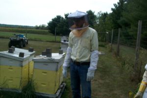
That’s where University of Wisconsin–Stout Biology Professor Jim Burritt steps in. Over the past couple of years, Burritt has led a study to find new ways to analyze the bees.
“The main goal for the study was to develop a tool–a tool that could be used by other people to evaluate the health of bees”, said Burritt, a beekeeper himself. The tools he and his students developed are similar to the techniques that doctors use for humans.
“If you get sick, one of the first things the doctor is likely to do is to collect a blood sample and look at the cells,” explained Burritt. “So, that is what we did for the bees. We learned how to collect their blood–it’s actually not called blood its called hemolymph–and we developed a new way to examine the cells in a way that had never been done.”
Bee hemolymph is like human blood but it is clear and simpler.
In order to examine the hemolymph, Burritt and his students used a microscope and a flow cytometer. Using a microscope to look at the cells at first seemed like a straightforward approach, but what they quickly discovered was that bee cells behave differently than human cells.
“We found that the cells won’t stick to a glass slide, and it seemed like nothing we would do would allow us to bind the cells to a slide so we could see them under a microscope,” Burritt said.
Luckily, Professor Randy Daughters had a solution. Burritt explained what Daughters said to do, “If we first coated the slide with a protein, with gelatin actually, then the cells would stick very well. Once they would stick, then we could stain them and look at them under the microscope.”
As of today, Burritt and the students have made thousands of bee cell smears on slides.
The flow cytometer that they used offered measurements on the cells and gave data on very large populations of cells, categorizing them into groups. What they found is that bees have at least four different types of cells. Burritt and the students then compared how the percentages of each cell type differ from bee to bee. It turns out that the hemolymph from bees in one hive can be drastically different than the hemolymph from bees in another. It is unclear what this means but further research is being done.
“We’re really trying to understand why bees have these changing profiles. That’s one thing we’re trying to do, “ said Burritt. “And the other is, we’re trying to associate these certain profiles with conditions of health or disease, or even time of year.”
The research being done here at Stout may lead to finding out why honeybees’ numbers are declining.
“Hopefully,” Burritt said, “we’ll be able to develop a better way to understand the things that are causing problems for bees, and there are a lot.”

